- 459 Hume Street Collingwood, ON, L9Y 1W9
- 705-445-2550

- Donate
Ultrasound
Diagnostic ultrasound is a non-invasive imaging test that uses high-frequency sound waves to create real-time images or video of organs and soft tissues in the body. Since these sound waves cannot pass through bone or air, ultrasound is commonly used for imaging soft tissues and fluid-filled spaces. This includes the liver, kidneys, gallbladder, reproductive organs, thyroid, scrotum, breast, blood vessels and obstetrics.
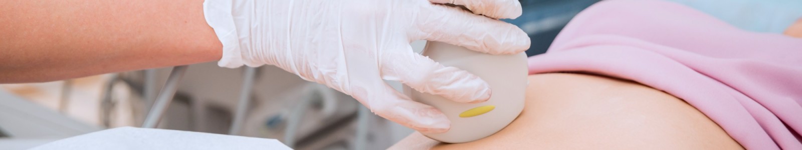
During the exam, a diagnostic medical sonographer (DMS) will place the transducer (a ultrasound probe) on your skin or, if needed, use a specialized probe for internal imaging. A thin layer of gel helps the sound waves travel smoothly from the probe through the gel and into your body. These sound waves bounce off internal structures and return to the probe, creating an image.
All images are interpreted by a radiologist and a detailed report is provided to your referring healthcare provider within a few days. Diagnostic medical sonographers cannot disclose results, including fetal sex.